What is Abdominal Separation During Pregnancy or Diastasis Recti?
What happens to your body during Pregnancy?
During Pregnancy, your body undergoes some incredible changes to accommodate a new-growing baby. One of them is the expansion of the muscles in your abdomen to make room for the baby.
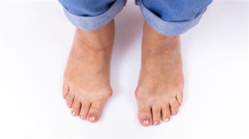
This results in a prominent bump on the side of the foot, which may cause pain, swelling, redness, and difficulty wearing particular shoes. There are many ways to prevent the development and slow the progression of a bunion. Today, we will be looking at management via exercise and foot strengthening.
Which Muscles Are Affected by Abdominal Separation During Pregnancy?
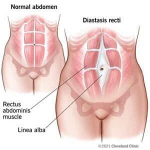
The rectus abdominis, which is the most superficial layer of your abdominal muscles, also referred to as your ‘six pack,’ is divided into a left and right half by a thick band of connective tissue called the linea alba. Throughout Pregnancy, the uterus expands to make room for the baby to grow, which can cause stretching across the rectus abdominis. The widening and weakening of the linea alba connective tissue creates a separation between the right and left sides of the rectus abdominus muscle. Once the baby is delivered, the linea alba can often heal independently due to its high elasticity. However, when the tissue loses its elasticity from being overstretched, the gap cannot close. Some separation is expected (1.5 cm-2 cm). However, any separation greater than 3cm is considered significant and is referred to as diastasis recti. This condition can be seen in both pregnant and postpartum women.
What are the Common signs and symptoms of Abdominal Separation?
- A visible bulge or ‘pooch’ that protrudes just above or below the belly button.
- Coning or doming when you contract your ab muscles.
- Pelvic or hip pain
- Lower back pain
- Poor posture
Rehabilitation Exercises for Abdominal Separation
Studies have shown that deep core stability exercises can reduce the severity, width, and occurrence of diastasis recti in pre-and post-natal periods. Exercises focusing on deep core muscles such as the pelvic floor, transverse abdominis, and obliques can help build strength and re-establish the connections between your abs and supporting muscles.
In a study that examined the effectiveness of a 12-week exercise program for those 6-24 months postpartum, a meaningful change in diastasis recti measurements was seen in two of the six locations along the linea alba. Exercises consisted of diaphragmatic breathing, bridge variations, plank variations, and deadbug variations.
A regular exercise routine has been shown to help and even heal diastasis recti symptoms. However, before you start, it is important to know which exercises are considered safe for you and which may aggravate your condition.
What can make Diastasis Recti Worse or Aggravate Them?
Certain activities can exacerbate Diastasis Recti, causing the abdominal muscles to separate, further hindering the healing process. Poor posture and heavy lifting can strain the abdominal muscles excessively, increasing intra-abdominal pressure and further separating the rectus abdominis muscle.
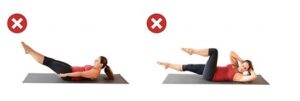
Exercise movements that can also put excessive strain on the abdominal muscles and that should be avoided include:
- Traditional Crunches or Sit Ups
- Reverse Crunches
- Planks or Push Ups (unless using modifications)
- Double Leg Lifts
- Any exercise that causes your abdominals to bulge, cone, or dome.
How can Physiotherapy & Exercise Physiology help people with Abdominal Separation?
Effectively managing diastasis recti requires an individualised approach. A Physiotherapist would be able to assist by assessing the area and diagnosing the Rectus Diastasis, from here Physiotherapists and Exercise Physiologists work together to incorporate targeted exercises, address core stability, and adopt a gradual progression.
A safe treatment plan would be focused around:
- Engagement of your Deep Core Muscles:
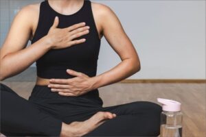
Focusing on exercises that engage and activate the deep core muscles, including the transverse abdominis and pelvic floor muscles, can help support the abdominal wall and promote the healing of the separation.
Incorporating diaphragmatic breathing (also known as belly or deep breathing) will also help enhance deep core activation.
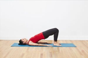
- Progressive Abdominal Strengthening:
Gentle exercises such as pelvic tilts and bridges will help activate the core. These exercises can then gradually progress to more challenging exercises that engage the abdominal muscles without causing excessive strain. Once foundational strength is established, modified or traditional exercises such as planks and crunches can be gradually incorporated to help further engage the deep core muscles while minimising intra-abdominal pressure.
- Pelvic Floor Rehabilitation:
Pelvic floor exercises will help to strengthen the muscles that support the pelvic organs and improve core stability.
- Whole-Body Conditioning:
Cardiovascular exercises and full-body strength training can enhance overall fitness.
Through an individualised and comprehensive approach, individuals may reduce symptoms, prevent diastasis recti complications, regain strength and function back in their abdominal muscles, and improve their overall quality of life.
References:
Chen, B., Zhao, X. and Hu, Y., 2023. Rehabilitations for maternal diastasis recti abdominis: An update on therapeutic directions. Heliyon.
Emma Boyes – Senior Physiotherapist (2024), WA Sports Med Physiotherapy Health Performance, Available at https://www.wasportsmed.com.au/blog/rectus-abdominis-diastasis-rad-management-and-training-plan (Accessed 18/04/2024)
Laframboise, F.C., Schlaff, R.A. and Baruth, M., 2021. Postpartum exercise intervention targeting diastasis recti abdominis. International journal of exercise science, 14(3), p.400.
Lyndsay Provencio, PT, DPT (2024), The Prehab Guys. Available at https://theprehabguys.com/what-you-need-to-know-about-diastasis-recti-abdominis-exercise/ (Accessed 18/04/2024)
Thabet, A.A. and Alshehri, M.A., 2019. Efficacy of deep core stability exercise program in postpartum women with diastasis recti abdominis: a randomised controlled trial. Journal of musculoskeletal & neuronal interactions, 19(1), p.62.